Point Of Care UltraSound & COVID-19
Daily thoughts & numbers on the COVID-19 pandemic from a M.D. working in the front lines at the Emergency Department in a hospital in Stockholm, Sweden.

POCUS with focus on lung ultrasound is a great additional diagnostic tool for us at the ER and ED to help diagnose and monitor the development of our patients with COVID-19 pneumonia.
First, look at the pleural line, and do it systematically.
It doesn’t matter what numeric zone standard you use, what matters is that you are consistent and systematic.
Once you start looking at the lungs apically in a healthy lung you’ll see the following:
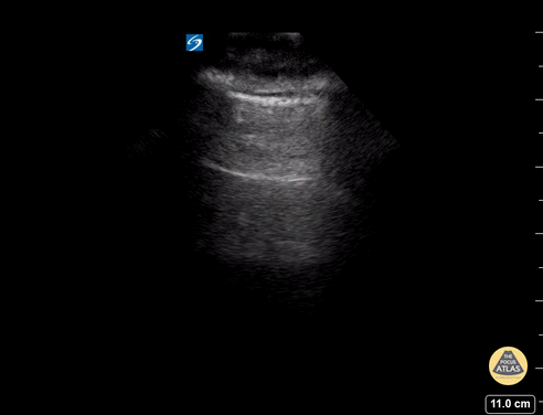
Image: healthy lung.
The pleural line is visualized at top of the image as a thin & regular white line (no sign of inflammation/infection), there is movement/lung sliding (no sign of pneumothorax), and there is a-lines (the second horizontal white line below the the top one – a sign of a “dry” lung”).
In a COVID-19 pneumonia what you’ll (sometimes) get is the following pleural line:

The pleural line above is irregular, there is a bump in the pleura (sign of a infection/subpleural consolidation), and b-lines following that bump (the moving vertical line, sign of a “wet” lung).

Image: day 5 after symptoms, in a patient developing a COVID-19 pneumonia.
Between the pleural line in the image to the left above and the thorax wall there is now fluid/edema shown as black (fluid becomes black with ultrasound). There are also several b-lines (signs of a “wet” lung, in other words: interstitial fluid) in each of the two captured images above, initially my experience is that these are seen more peripherally in some patients while they can maintain a-lines central/apical (COVID-19 seems, radiologically, to affect the lungs more peripheral/lower parts/subpleural *).

Image: pleural findings in a more advanced COVID-19 pneumonia.
Presumably a (radiologically) more severe pneumonia seen in the image above, with shredding (a highly irregular/inconsistent pleural line), several bumps indicative of infection/subpleural consolidation, and more prouncanced amount of fluid/edema between the pleural line and the thoracic wall.
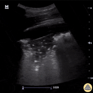
Image: plural findings in a patient with bacterial pneumonia.
The patients with COVID-19 can also get a secondary bacterial infection as an addition to their COVID-19 pneumonia. In the image above signs of a larger subpleural irregularity is seen in the middle of the pleura line with consolidation (signs of a collapsed piece of lung) and within it: static air bronchograms (air-filled bronchi (dark) being made visible by the opacification of surrounding alveoli (grey/white), it is almost always caused by a pathologic airspace/alveolar process, in which something other than air fills the alveoli *). Keep in mind that static airbronchograms do not rule in bacterial pneumonia, they can also be a feature of just a COVID-19 pneumonia – a sign of resorptive atelectasis. You’ll need to look at the clinic and lab results for additional guidance as (almost) always with POCUS.
The COVID-19 pneumonia does not seem to produce large amounts of pleural effusion, however small amounts can be seen, sometimes also alongside with posterior/diafragmal/lower lung b-lines.
Sources:
- [SWE] Powerpoint on COVID-19 & POCUS
- The POCUS Atlas Images – COVID-19 Ultrasound Findings
- [Study 2020] Lung Ultrasound in Emergency and Critically Ill Patients – Number of Supervised Exams to Reach Basic Competence
- [STUDY 2020] Findings of lung ultrasonography of novel corona virus pneumonia during the 2019–2020 epidemic
- Radiopaedia on COVID-19
COVID-19 Numbers Sweden 2020-04-08 *
- 687 deaths nationally
- 419 of them in Stockholm
- 8 419 confirmed cases nationally
DEATHS [SWE]
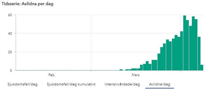
New confirmed deaths daily in Sweden.
(Number of COVID-19 cases over time in Sweden, updates during mornings at 14:00 so todays numbers are not yet complete until the next day).
ICU [SWE]
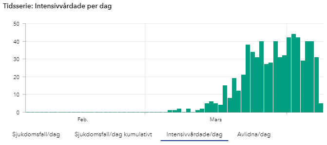
New Covid-19 patients Being Treated at the ICU in Sweden.
(Number of COVID-19 cases over time in Sweden, updates during mornings at 14:00 so todays numbers are not yet complete until the next day).
NEW CONFIRMED CASES [SWE]

New confirmed cases daily in Sweden.
(Number of COVID-19 cases over time in Sweden, updates during mornings at 14:00 so todays numbers are not yet complete until the next day).
TOTAL CONFIRMED CASES [SWE]
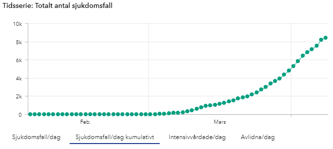
New confirmed cases accumulated over time in Sweden.
(Number of COVID-19 cases over time in Sweden, updates during mornings at 14:00 so todays numbers are not yet complete until the next day).
- Intensive care Sweden: *
- 681 COVID-19 confirmed patients treated in the ICU in total so far
- 10,2 days from symptoms to ICU
- 24,4 % women

Total number of recorded of COVID-19 patients needing ICU care each week. From Week 10 it seems to more than triple each week for the first 4 weeks. From week ten: 5 → 24 → 130 → 420 → 743 → (856 this week up until now, however since it’s only wednesday this weeks numbers are yet incomplete)
COVID-19 Numbers Globally (updated today 2020-04-08 at 14:00 CET) *
- 79 385 confirmed deaths
- 1 356 780 confirmed cases
- 6 695 new deaths, daily change: 33,37%
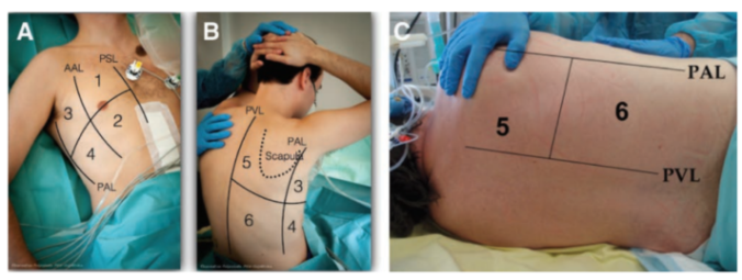
Image: Denver Health & The POCUS Altas on COVID-19 findings with lung ultrasound *

That’s it until tomorrow, keep calm and stay safe!
